
Gynecology
We take special care in addressing each woman’s specific needs and concerns, and place an emphasis on educating our patients so they feel very comfortable with their situation, expectations, and the processes involved.
IN-OFFICE PROCEDURES:
In-Office procedures take place at Northside Women’s Health, located at 550 S. Cleveland Avenue, Suite D, Westerville, OH 43081
Colposcopy
A colposcopy is a procedure that we do in the office to look closely at the cervix. Any abnormal areas will be biopsied. The procedure typically takes about 5 to 10 minutes.
First we place a speculum in the vagina and then we wash the cervix off with two types of stains, and then we use a magnifying glass, called a colposcope to closely examine the cervix. Any areas that appear abnormal we will biopsy. If there is any bleeding from the biopsy site we place a solution to help stop the bleeding. The solution may drain from the vagina and it can be a yellowish color or it can be a brownish blackish color. The biopsy results are typically back within a week.
Endometrial Biopsy
An endometrial biopsy is a procedure that is done in the office to obtain a sample of the endometrium, the lining of the uterus. We typically recommend that you take some ibuprofen or Advil prior to the procedure. We may also prescribe medicine, misoprostol, to be placed in the vagina the night before and the morning of the procedure to soften the cervix.
First we do an examination of the uterus to determine the position of the uterus. Then we place a speculum in the vagina. We wash the cervix and then we use a very small straw like biopsy device, called a pipelle, and insert it through the cervix up into the uterus. We apply a small amount of suction and obtain some of the endometrial tissue of the uterus, and then place it into a specimen cup for the pathologist to examine. When we place the pipelle, or small straw through the cervix you typically have a cramp or contraction. The cramping typically resolves within a few minutes. The biopsy results are usually available in about one week.
SIS
An SIS, or saline infusion sonogram, is a special type of ultrasound that is done in the office. In this ultrasound, saline solution is placed inside the uterus, which allows us to see the contour of the uterine cavity. This can help us see if there are any masses inside the uterus, such as a fibroid or a polyp. This ultrasound procedure usually takes 5 to 10 minutes. You may have some mild cramping, that is typically well-managed with ibuprofen
We will first do a pelvic examination to determine the size and position of your uterus. We will place the speculum in the vagina and wash the cervix with some soap. We then place a very small straw inside the uterus and attach a syringe of Saline. We remove the speculum from the vagina and the ultrasonographer places the ultrasound probe or camera inside the vagina. We instill saline inside the uterus and the sonographer takes pictures. You’ll be able to see the images on the screen while this is being done.
We then remove the catheter from the cervix and review the images with you. You will have watery blood tinged discharge for the next couple of hours as the the saline extrudes from the uterus.

PHOTO FROM THE 24HOUR MOMMY
Endosee
An Endosee, is an in office procedure, that allows us to look inside the uterus. We can also take a sample of the uterine tissue. The procedure, usually takes about 15 minutes. You’ll have some cramping and we recommend that you take ibuprofen before the procedure.
We will examine your uterus and determine the shape and position of the uterus. We place a speculum in the vagina, we wash the cervix with soap, and we slide a very thin camera inside the uterus. We then attach a device that looks similar to a telephone. We instill the uterus with saline, and we are able to see the inside of the uterine cavity. We can see the tubal ostia, or the openings to your fallopian tubes. We also can determine if there are polyps or fibroids or excessive tissue. Occasionally we utilize this procedure to remove an IUD if the strings have retracted into the uterus.
We take the camera out of the uterus and then we obtain a sample of the tissue and send it to the pathologist for examination.
After the procedure you’ll have bloody watery discharge draining from the uterus. We will review the pictures and the video with you immediately after the procedure.
Vulvar Biopsy
A vulvar biopsy is a procedure typically done in the office setting to test for vulvar cancer or other skin diseases. The vulva is the outer part of a woman's genitals. We may perform this procedure if there are areas that appear abnormal or conditions that are not responding to treatment. The procedure involves numbing the area with a cream and/or shot, then removing a small piece of tissue, and applying a solution to help the area heal. Sometimes, there will be a stitch placed to reduce any bleeding. The tissue is then sent to a lab for testing. The procedure usually takes less than 10 minutes.
After the procedure, we may ask that you wear loose clothing and underwear to allow for appropriate healing. We will usually see you 1-2 weeks after for follow-up.
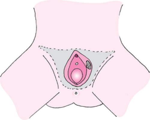
IMAGE FROM OBGYN KEY
LEEP (Loop Electrosurgical Excision Procedure)
LEEP is a treatment to remove precancerous cells from the cervix. Removing precancerous cells helps stop them from developing into cervical cancer. This is a procedure performed most commonly in the office under local anesthesia and typically takes less than 15 minutes to do. We place a speculum in the vagina in the same way as for a Pap smear and numb the cervix with Lidocaine. The procedure uses a small wire loop attached to an electrical current. When the loop is passed over cervical tissue, it cuts away a layer of abnormal cells. The removed tissue is sent to a lab for testing. At the end of the procedure, we apply medicine on the cervix to help with any bleeding at the cervix. It is normal to feel some pressure, dull ache or cramping. Sometimes, you may feel your heart race due to the numbing medication.
A LEEP should be done when you are not having your period to give a better view of the cervix. You may have thick brown discharge after the procedure for a week and watery discharge for up to a week after that. Occasionally the discharge may have a strong odor to it that is normal. We ask that you refrain from intercourse or placing anything in the vagina for 2 weeks to allow the cervix to heal. The results of the biopsy from the tissue removed will usually return in less than 2 weeks. We will see you for a followup appointment in 1 month.
IUD
IUD, stands for intrauterine device. We insert an IUD into the uterus for long acting reversible contraception, or to treat heavy bleeding, or to treat painful menstrual cycles. IUD Placement typically takes about 5 to 10 minutes.
We typically ask the patient to place two tablets of Misoprostol in the vagina the night before their procedure, and two tablets in the vagina the morning of the procedure. This helps soften the cervix. We also recommend taking ibuprofen 30 minutes prior to the appointment to help reduce the cramping associated with IUD placement.
The patient first gives a urine sample for a pregnancy test prior to IUD placement. Then the clinician feels the position of the uterus. The speculum is placed in the vagina, and the cervix is washed with a cleaning solution. Under ultrasound guidance an instrument called a sound is placed through the cervix to the top of the uterus. This allows us to measure the length of the uterus. We set the measurement on the IUD device and insert the IUD while watching on the ultrasound. We place the IUD at the very top of the uterus, called the fundus. We then cut the strings of the IUD so they are about 2 cm long, and remove the speculum from the vagina.
We will schedule a follow up appointment in a month to do an ultrasound or an examination to ensure that the IUD is in the correct position in the uterus.
You will likely have cramping the day the IUD is placed and perhaps a couple days afterwards. This is typically well-managed with ibuprofen. For the two months following IUD placement, you can expect to have irregular bleeding and spotting. This is normal.
The ParaGard, copper IUD is effective immediately. The hormone releasing IUDs such as the liletta, kylena, and Mirena, are also effective immediately if placed within seven days of the start of your menstrual cycle. If placement was not within seven days of the start of your period, you should abstain from intercourse or use a back up method for seven additional days.
We will schedule a follow up ultrasound or pelvic exam in a month to ensure that the IUD is in the correct position in your uterus.
We will submit your IUD information to your insurance to determine if you have IUD coverage and will call you back to let you know and will schedule your appointment at that time.
We place the IUD under ultrasound guidance. Some insurance companies cover the Ultrasound and some do not. If your insurance company doesn’t cover the Ultrasound, you will be responsible for that payment.
Please remember - No unprotected intercourse for two weeks prior to your IUD placement. Effective contraception is considered condoms all of the time, birth control pills, or patches, rings, or a current IUD.
We utilize misoprostol, to soften the cervix prior to your IUD placement. We use this for most patients, some patients who have had a recent vaginal delivery, do not need to utilize this. Your clinician will determine if misoprostol is necessary or not. Please place two tablets of misoprostol in your vagina the night before your procedure, and two tablets of misoprostol in the vagina the morning of your procedure. This is to soften the cervix and make your IUD placement more comfortable for you. Occasionally people will have some cramps with the misoprostol, please just take ibuprofen as needed.
Please take 600 mg of ibuprofen about 30 minutes prior to your appointment.
Please eat something prior to your appointment.
When you arrive at the office to check in for your appointment, please come back and leave a urine sample. We do a pregnancy test prior to every IUD placement.
IUD INSTRUCTIONS

NEXPLANON® Insertion and Removal
NEXPLANON® is a highly effective contraceptive device which contains a low dose of hormone to prevent pregnancy for up to 3 years. NEXPLANON is placed in the inner, upper arm in a healthcare provider’s office. The flexible device is about 1½ inches long and approximately 1/8 inch in diameter. Women may have no period, a monthly period, or irregular bleeding caused by the hormone in NEXPLANON.
While your NEXPLANON is inserted, you will lie down and place your arm comfortably near your head. The healthcare provider may make two marks on your skin to identify where NEXPLANON will be placed. Your skin will be cleaned with betadine and numbing medication will be placed under the skin. Most women would describe this as a small stick and stinging feeling. After your arm is numb, an insertion needle containing NEXPLANON is placed beneath the skin. This insertion device is removed and the NEXPLANON remains in the arm. If you touch your arm, you can feel the device beneath the skin.
You will be asked to keep a tight bandage on for 24 hours and a smaller bandage for 3-5 days. You will be protected from pregnancy after 7 days. You can remove NEXPLANON at any time but it should be removed after 3 years.
NEXPLANON is typically removed in a healthcare provider’s office. Your skin will be cleaned with medical soap and numbing medication will be placed in your arm. This medication feels like a stick and sting and will prevent pain while your NEXPLANON is removed. A small incision will be made in your skin and NEXPLANON will be removed through this incision. If you are continuing to use NEXPLANON, a new device will be placed through the same incision. The incision will be covered with a special bandage.
You are immediately able to get pregnant after NEXPLANON is removed.

IMAGE CREDIT: NEXPLANON®
SURGICAL PROCEDURES:
Surgical procedures take place at:
Mount Carmel St Ann’s Hospital (500 South Cleveland Ave, Westerville, OH 43081)
Dublin Surgery Center (5005 Parkcenter Ave, Dublin, OH 43017)
Endometrial Ablation
Endometrial ablation is a procedure used to burn the lining of the uterus, or the endometrium. For most women, menstrual bleeding is successfully reduced or stopped within one year of the procedure. Prior to the procedure, a sample of the uterine lining, or endometrial biopsy, is performed in the office to ensure there are no abnormal or precancerous cells causing the heaving bleeding.
Endometrial ablation is a hospital based procedure. The cervix is dilated and typically hysteroscopy is performed prior to the procedure to examine the uterine cavity prior to the ablation. The ablation device is inserted into the uterus and using radiofrequency energy the lining is burned.
You are typically in the recovery room for about an hour, and then you are discharged to home and someone will need to drive you. We usually recommend ibuprofen and acetaminophen in an alternating manner, to control any cramping or pain. It is common to have dark brown or watery discharge following the procedure. You should not have anything in the vagina until you are seen at your follow up appointment by your clinician to make sure that the cervix is closed. We will review your pathology report and any pictures taken during your surgery.
Hysteroscopy
Hysteroscopy is a procedure performed is a hospital based procedure in which we use a small camera to visualize the cervical canal and endometrial lining. Your cervix is dilated and a small camera is inserted into the uterus and we examine the size and shape of the uterine cavity. We do this to see if there are any fibroids (benign muscle tumors), or polyps, placental fragments, or scarring. IV fluid is used to distend the cavity. Polyps and fibroids in the uterine cavity can be removed and sent to the pathologist for evaluation.
You are typically in the recovery room for about an hour, and then you are discharged to home and someone will need to drive you. We usually recommend ibuprofen and acetaminophen in an alternating manner, to control any cramping or pain. You should not have anything in the vagina until you are seen at your follow up appointment by your clinician to make sure that the cervix is closed. We will review your pathology report and any pictures taken during your surgery.

IMAGE FROM HEALTHYWA
Hysteroscopic Polypectomy
Uterine polyps are a common cause of heavy periods and intermenstrual bleeding. In most cases, these are benign overgrowths of tissue. A polypectomy, or removal of the polyp, can be performed during hysteroscopy. Hysteroscopy is a procedure in which your cervix is dilated and a small camera is inserted into the uterus. A device is inserted through the camera and the polyp is removed. The polyp is sent to the pathologist for evaluation.
You are typically in the recovery room for about an hour, and then you are discharged to home and someone will need to drive you. We usually recommend ibuprofen and acetaminophen in an alternating manner, to control any cramping or pain. You should not have anything in the vagina until you are seen at your follow up appointment by your clinician to make sure that the cervix is closed. We will review your pathology report and any pictures taken during your surgery.

IMAGE FROM THE TORONTO VIDEO ATLAS OF SURGERY
Hysteroscopic Myomectomy
Uterine fibroids are a common cause of heavy periods and intermenstrual bleeding. In most cases, these are benign overgrowths of tissue. A submucosal fibroid is located under the uterine lining. A myomectomy, or removal of the submucosal fibroid, can be performed during hysteroscopy. Hysteroscopy is a procedure in which your cervix is dilated and a small camera is inserted into the uterus. A device is inserted through the camera and the fibroid is removed. The tissue is sent to the pathologist for evaluation.
You are typically in the recovery room for about an hour, and then you are discharged to home and someone will need to drive you. We usually recommend ibuprofen and acetaminophen in an alternating manner, to control any cramping or pain. You should not have anything in the vagina until you are seen at your follow up appointment by your clinician to make sure that the cervix is closed. We will review your pathology report and any pictures taken during your surgery.

IMAGE FROM THE TORONTO VIDEO ATLAS OF SURGERY
Laparoscopy
Laparoscopy is a hospital based procedure used to evaluate the pelvis and operate with minimal incisions. After receiving general anesthesia, a small port is inserted near your belly button and the abdominal cavity is inflated with carbon dioxide gas. A camera is inserted into the port which allows your doctor to look at your uterus, tubes and ovaries and pelvic side wall. Most of the time, a small instrument is inserted into your uterus so that the doctor can visualize the area underneath your uterus. Small accessory ports are typically placed by each hip bone to provide access for surgical instruments.
Operative laparoscopy could be used to remove fibroids (myomectomy) or operate on ovaries (remove a cyst or the entire ovary), fallopian tubes, remove ectopic pregnancies or endometriosis. A hysterectomy can also be performed laparoscopically. All the tissues removed are sent to the pathologist for examination.
After the procedure, you are in the recovery room for 1-2 hours. You will need someone to drive you home that day. You may experience abdominal pain and bloating related to the carbon dioxide gas. Gas-ex can alleviate this symptom. We recommend alternating Tylenol and motrin for pain. Laparoscopy typically involved a 4 week recovery time from intense physical activity. You will be seen in the office in 1-2 weeks from your procedure to evaluate your incisions and discuss the findings of the surgery.
Hysterectomy
Hysterectomy is surgery to remove the uterus. It is major surgery and is used to treat many women's health conditions such as uterine fibroids, pelvic pain, abnormal uterine bleeding, uterine prolapse, and gynecologic cancer. There are different ways to perform a hysterectomy, including the abdominal route (making a large incision in the abdomen), laparoscopic route (a few small incisions in the abdomen), and vaginal (an incision at the top of the vagina).
The route of hysterectomy may depend on the reason for surgery, size of the uterus, and the patient's surgical history. It may also affect the patient's time of recovery. Hysterectomy does not always mean that we take out the ovaries or cervix. It is important that you discuss the route of surgery and specific organs removed with your doctor.

IMAGE FROM NATIONAL WOMEN’S HEALTH NETWORK
Dilation and Curettage
A dilation and curettage, or a D&C, is a hospital based procedure where we take a thorough sample of the entire uterine lining. We typically also do a hysteroscopy, meaning we place a camera inside the uterus and examine the size and shape of the uterine cavity. We do this to see if there are any fibroids (benign muscle tumors), or polyps, placental fragments, or scarring. We then do a curettage, which means we gently remove the uterine lining. This tissue is then sent to the pathologist for evaluation.
You are typically in the recovery room for about an hour, and then you are discharged to home and someone will need to drive you. We usually recommend ibuprofen and acetaminophen in an alternating manner, to control any cramping or pain. You should not have anything in the vagina until you are seen at your follow up appointment by your clinician to make sure that the cervix is closed. We will review your pathology report and any pictures taken during your surgery.

IMAGE FROM THE MAYO FOUNDATION
Abdominal Myomectomy
Abdominal myomectomy, also known as "open myomectomy," is a major surgery that removes benign masses called fibroids from the uterus. It is performed in a hospital under general anesthesia. An incision is made on the lower abdomen and the fibroids are cut out from the wall of the uterus. The uterus is then sewn back up in layers of stitches. The tissue removed will be sent to a lab to ensure that the masses removed are benign fibroids. The typical hospital stay is about 2 days after the surgery. We usually recommend no heavy lifting or strenuous activity for 6 weeks after the surgery as the incision is healing. We will usually see you for follow-up 1 week and 6 weeks after surgery.
